
The Use of Confocal Laser Scanning
Microscopy and a Novel Imaging Program
for Quantifying Dentin Tubule Occlusion
Reading time: 2 minutes
Author: Deon Hines, PhD
The use of confocal laser scanning microscopy and a novel imaging program for quantifying dentin tubule occlusion
Colgate-Palmolive Company Technology Center, 909 River Road, Piscataway, New Jersey, USA
Poster#0742 2018 AADR/CADR 47th Annual Meeting Fort Lauderdale, Fla., USA
Study objectives
The purpose of this study was to investigate the in vitro dentin occlusion, utilizing a Confocal Laser Scanning Microscope, of an 8% arginine and calcium carbonate toothpaste (Colgate® Sensitive Pro-Relief) and two calcium phosphosilicate toothpastes (Sendoyne Repair & Protect). A novel imaging analysis method was utilized to quantify the amount of dentin occlusion.
Trial conditions and methods
Products under investigation
Test dentifrice 1: Colgate Sensitive Pro-Relief
(CSPR; Colgate-Palmolive Company, New York, NY)
Test dentifrice 2: Sensodyne Repair and Protect
(Novamin/Sodium Monofluorophosphate; GlaxoSmithKline, Brentford, London, UK)
Test dentifrice 3: Sensodyne Repair and Protect
(Novamin/Sodium Fluoride; GlaxoSmithKline, Brentford, London, UK)
Methods
Cut dentin specimens were polished, acid etched, dried and imaged. The dentin surface was treated with slurries by mixing 1 part PBS to 3 parts toothpaste for 30 seconds and subsequently rinsed and dried. The procedure was repeated 5 times. Percent occlusion was quantified based on the total scanned image area of open tubules before treatment versus the area of the existing open dentin tubules after treatment. The analysis of variance test was used to compare the mean percent occlusion for each toothpaste. A subsequent Tukey multiple comparison test was performed to assess pairwise comparisons of the toothpastes. A p-value < 0.05 indicated statistically significant differences among the samples.
Results
The percent occlusion for the three toothpaste samples was calculated to be 88% for CSPR, 72% for Novamin/Sodium Monofluorophosphate, and 50% for Novamin/Sodium Fluoride. The Tukey pair-wise comparison test shows statistically better (p< 0.05) occlusion for CSPR versus both the Novamin toothpastes. Additionally, the Novamin/ Sodium Monofluorophosphate provided statistically better (p< 0.05) occlusion than the Novamin/Sodium Fluoride toothpaste.
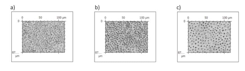
Confocal Images Used to Quantitatively Determine % Occlusion
Figure 1. Before treatment images of a) CSPR, b) Novamin/Sodium Monofluorophosphate, and C) Novamin/Sodium Fluoride
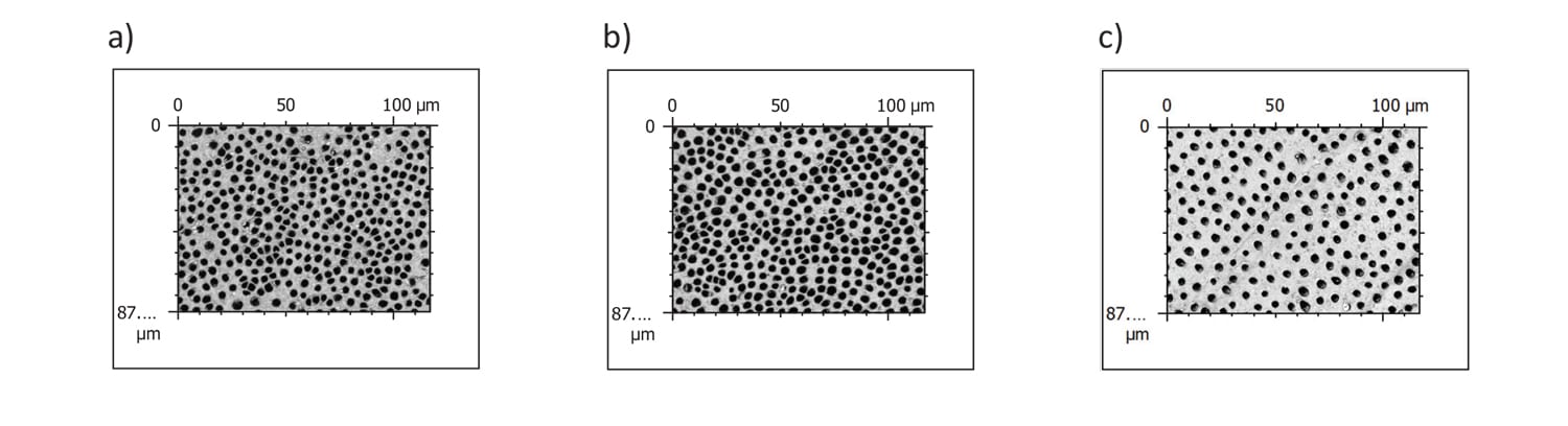
Figure 2. After treatment images of a) CSPR, b) Novamin/Sodium Monofluorophosphate, and C) Novamin/Sodium Fluoride
Conclusion
The results from this in vitro study and the imaging method indicate that treatment of dentin specimens with CSPR toothpaste exhibit significantly greater occlusion when compared to both the Novamin/Sodium Monofluorophosphate and Novamin/Sodium Fluoride. Novamin/Sodium Fluoride was significantly less effective at occlusion than Novamin/Sodium Monofluorophosphate.
